Learning Schedule and Objectives
| Week | Topics | Assigned Reading |
| 2 | Brain and cranial nerves | Ch. 8, 206-219; 223-228. |
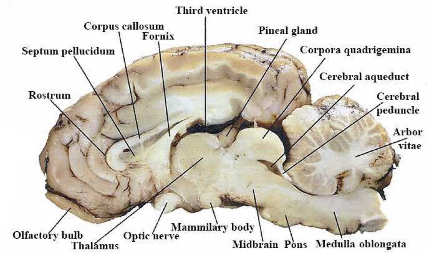
Dissect sheep brain and identify:
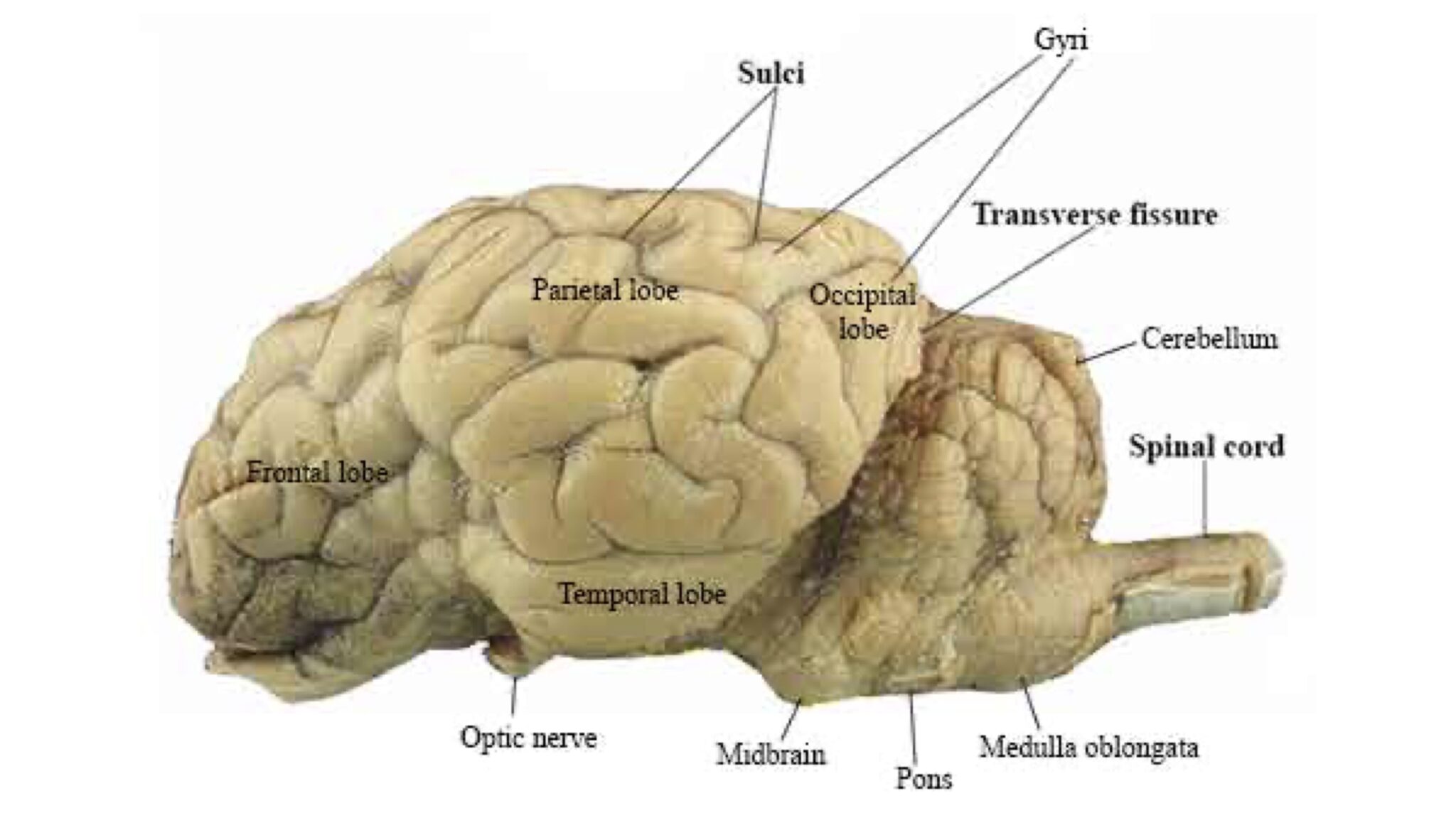
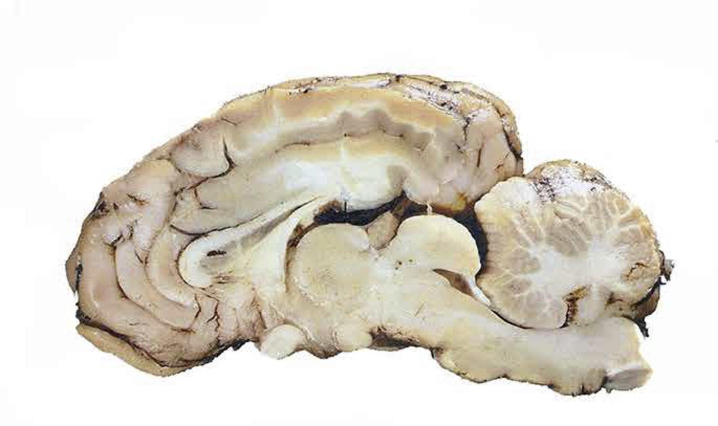
Cerebrum and cerebral Cortex, Dura matter, Ventricles, Pons, Medulla Oblongata, Cerebellum, Arbor vitae
Frontal, parietal, and occipital lobes, Thalamus, Pituitary, Hypothalamus, Gray matter and white matter
Brainstem, Olfactory bulbs, Optic nerves and optic chiasma
Identify the same on the brain model:
On the brain model identify all 12 cranial nerves
On the mink, be able to identify the following cranial nerves: I, II, VII, VIII, X
Important Notice
- Bring the Laboratory Manual to every Lab class. Preview manual before lab.
- Dissection Kit must be purchased and brought to lab starts from Lab1.
- Shoes with open tows and slippers do not allowed in the Laboratory Room.
- Read Biology Laboratory Safety guidelines or download Biology Lab Safety PPT.
- Above rules may not apply to remote learning, please reach out to your instructor.
- No makeup practical exam.
Slides
Download Lab2 PPT or View below
SCB209Lab-2Videos
Introduction to the Central Nervous System
Brain Model Walk-through, watch more: Brain model Internal and External view.
Sheep brain dissection video, watch more: Sheep Brain Dissection with labels
Lab Activities
Slide left or right to learn the labels of models.
- Brain Model – Lateral View
- Brain Model – Medial View
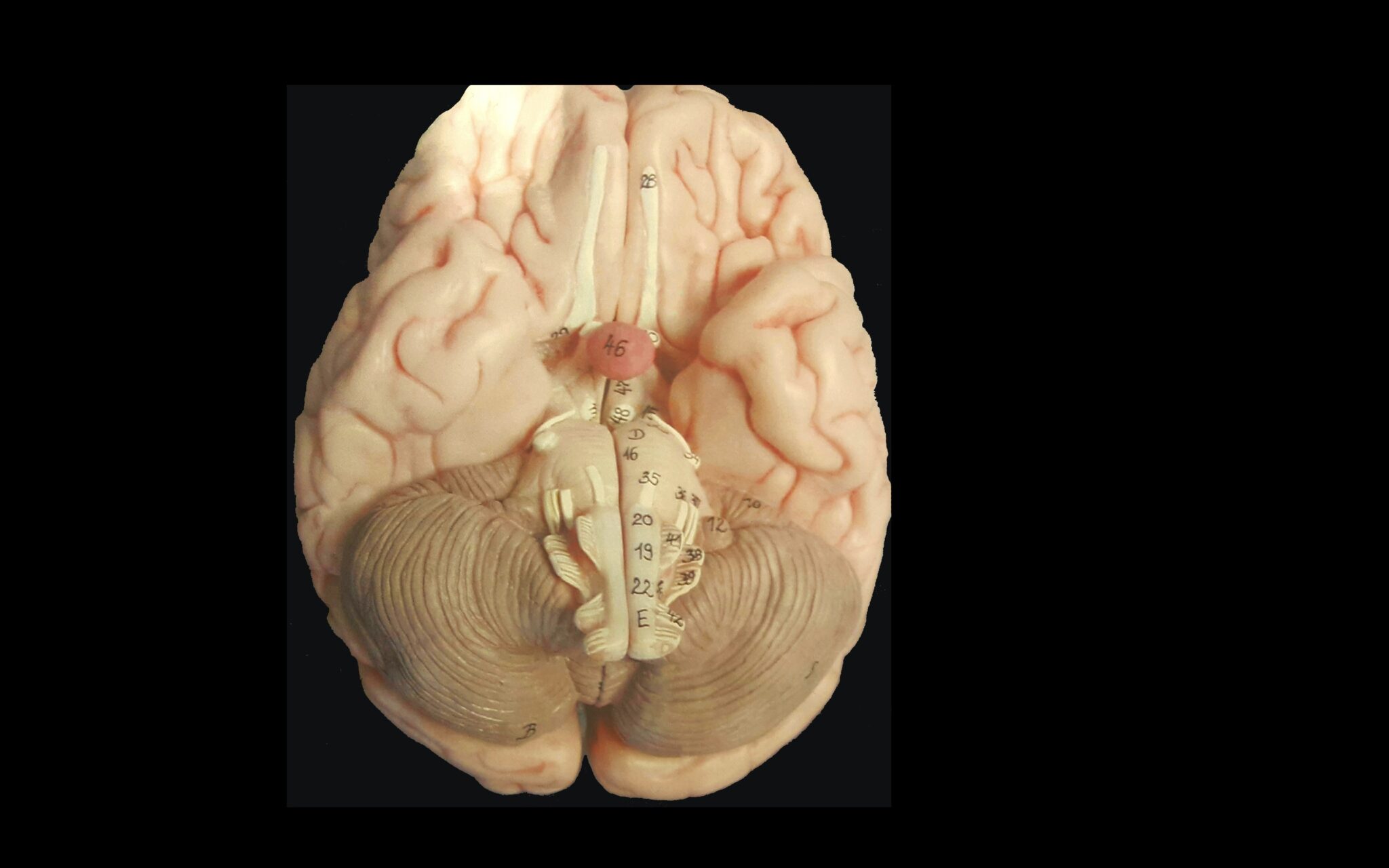
- Brain Model – Ventral View
- Brain Model – Cranial Nerves
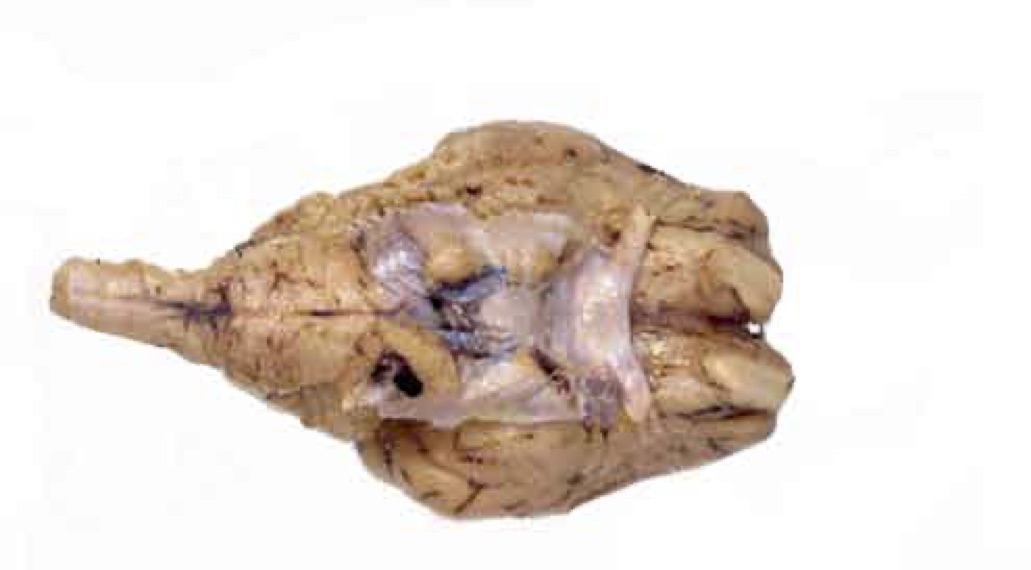
- Sheep Brain Specimen – Dorsal View
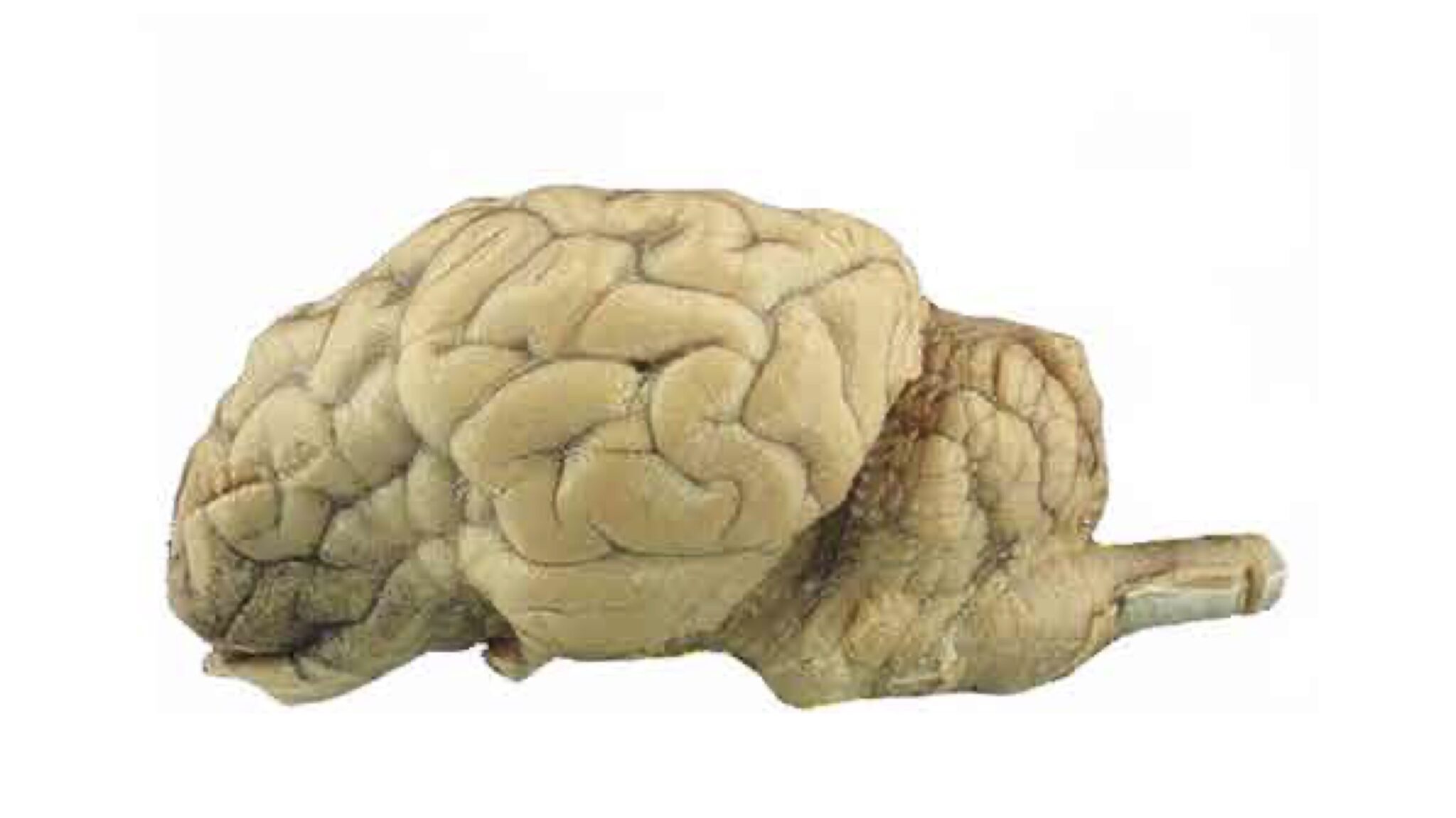
- Sheep Brain Specimen – Lateral View
- Sheep Brain Specimen – Sagittal View
- Sheep Brain Specimen – Ventral View
- Cranial Nerves Summery (Extra reading and video)
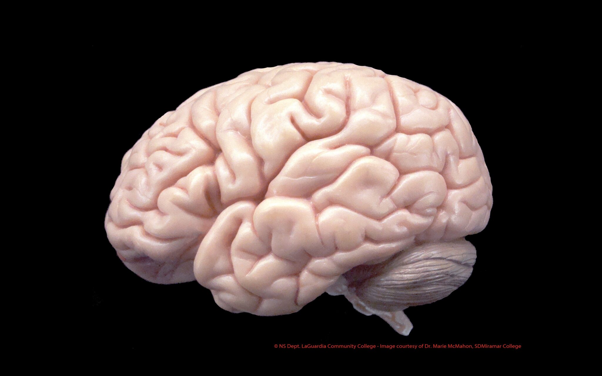
Brain Model – Lateral View

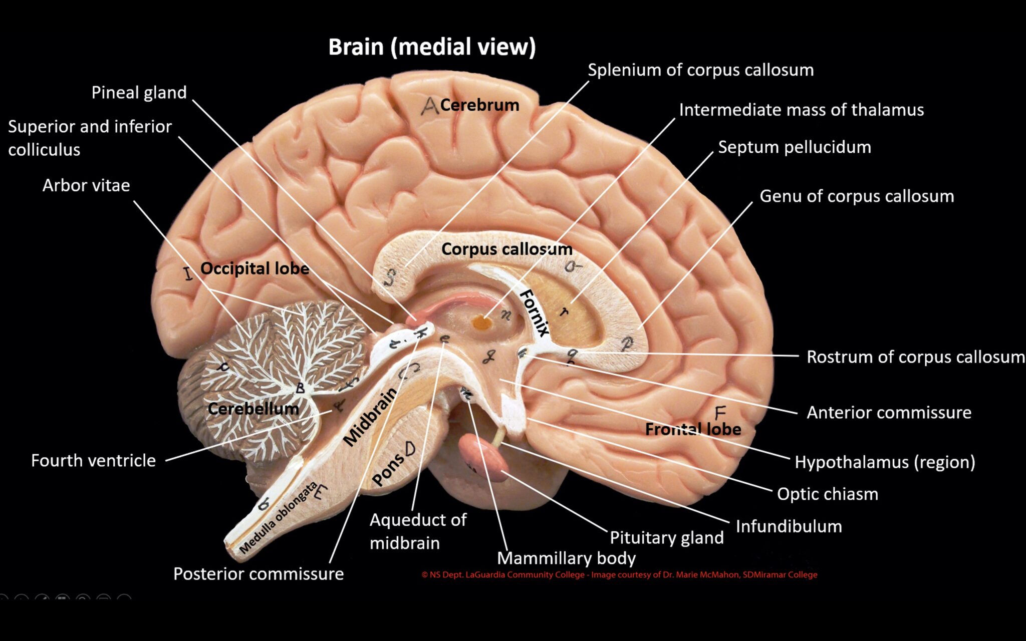
Brain Model – Medial View

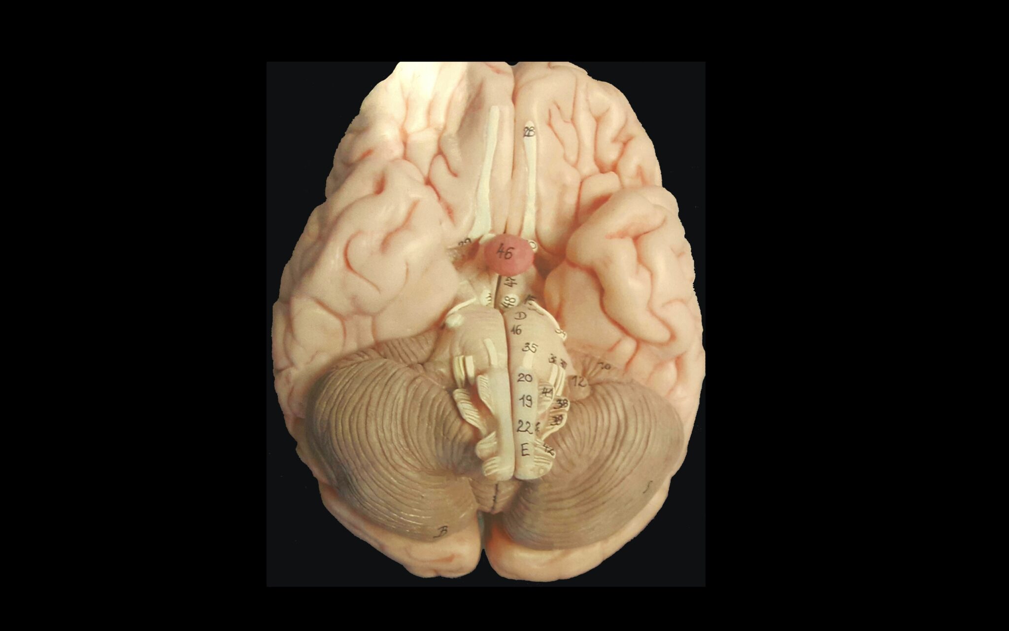
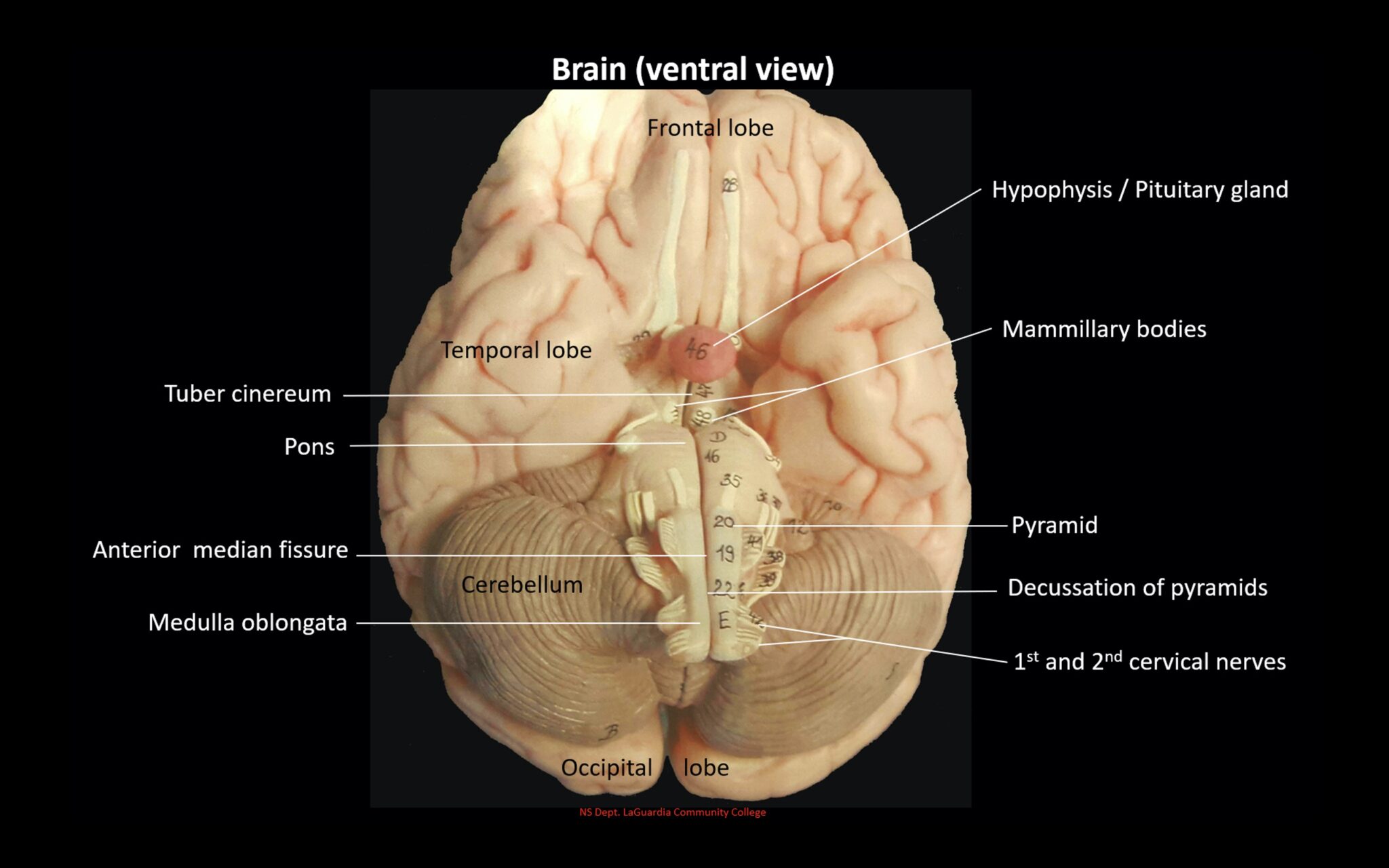
Brain Model – Ventral View
Brain Model – Cranial Nerves

Sheep Brain Specimen – Dorsal View


Sheep Brain Specimen – Lateral View
Sheep Brain Specimen – Sagittal View
Sheep Brain Specimen – Ventral View

Cranial Nerves Summery, Click here for 3D Anatomy Tutorial Video.
